Research

In the Field: Macro Photography, Microscopy & How Butterflies Create Color

YouTubeCompilation_All_Wings9.jpg

Working in the Amazon rainforest has its challenges. To name an obvious one, it can be difficult to take equipment into remote field sites in order to conduct research. Fortunately, we live in an exciting time, as technology is rapidly becoming simultaneously cheaper and more portable. In this post, I want to share a couple tools that I use to document small organisms in the rainforest, including the wing structures of butterflies and moths, as well as discuss the fascinating ways that biology creates color.

Macro shot of a Heliconius butterfly wing. The different colors (oranges, yellows, blacks) are caused by pigment production in each individual scale. MP-E65mm, ƒ/11.0, 1/125, ISO 200.

Macro shot of a Heliconius butterfly wing. The different colors (oranges, yellows, blacks) are caused by pigment production in each individual scale. MP-E65mm, ƒ/11.0, 1/125, ISO 200.

For starters, digital SLR cameras and macro lenses are powerful handheld tools that I use to document the biological diversity of tiny creatures that inhabit South America. I'm currently using a Canon 70D camera body equipped with the shockingly powerful MP-E 65 Macro lens. This lens is truly a macro beast, magnifying up to 5 times (aka a magnification ratio of 5:1) and allows me to get sharp images of microscopic structures, such as butterfly wing scales. For shots of the whole organism, I typically use the Canon EF 100mm f/2.8L macro lens, which I really love for its versatility and sharpness. In combination, the 100mm and MP-E 65 are a fantastic combination for macro photography in the field, allowing me to document small organisms such as insects, as well as zoom-in even closer to resolve  specific regions.

Here's a video explaining how these butterflies create color & using the Foldscope to investigate scale structures.

At our remote outpost in Sumaco, Ecuador, tinkering with my camera to photograph insects and butterfly wing scales.

At our remote outpost in Sumaco, Ecuador, tinkering with my camera to photograph insects and butterfly wing scales.

s an entomologist in the Amazon, I've been able to study a broad range of fascinating creatures, from Glowing Worms to Tentacled Caterpillars. More recently, I've become enthralled by the wings of butterflies and moths, and more specifically am curious about how these organisms produce such an incredible array of colors.

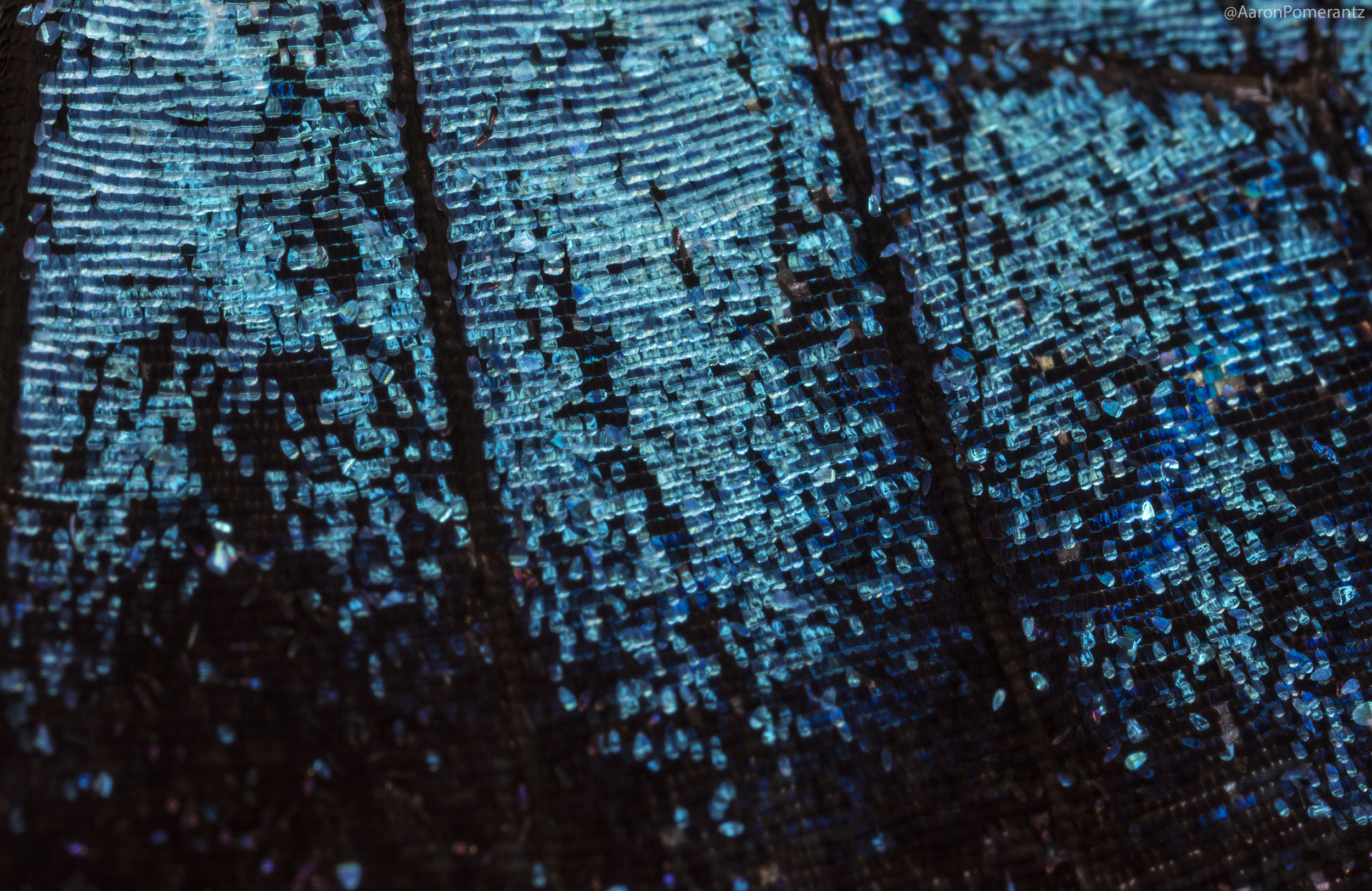
Macro of a Morpho wing, note the blue scales which do not contain any blue pigment. They contain nanostructures that bounce light back at the blue wavelength, a form of 'structural color'.

Macro of a Morpho wing, note the blue scales which do not contain any blue pigment. They contain nanostructures that bounce light back at the blue wavelength, a form of 'structural color'.

Butterflies and moths belong to the order Lepidoptera and all members have scales covering their bodies and wings (in Latin, lepis means scale and ptera means wing). With over 180,000 described species, the Lepidoptera are not only diverse in their numbers but also in their colors. Their color arises due to the nature of the scales that they produce and can be due to pigmentation as well as structural color. Whatever the origin, color results from an interaction between light and matter.

Like beautiful painted tiles, the scales on this Phantom butterfly range from shades of pink to entirely transparent. MP-E65mm, ƒ/11.0, 1/125, ISO 200.

Like beautiful painted tiles, the scales on this Phantom butterfly range from shades of pink to entirely transparent. MP-E65mm, ƒ/11.0, 1/125, ISO 200.

Owl Butterflies mating. The large eye spot on the hindwing is thought to startle potential predators like birds, a form of Batesian mimicry in which a harmless organism acquires protection by resembling a threatening animal. 

Owl Butterflies mating. The large eye spot on the hindwing is thought to startle potential predators like birds, a form of Batesian mimicry in which a harmless organism acquires protection by resembling a threatening animal. 

But even with the best macro lenses, it's still tough to resolve the scale structures on the wings of these insects. To get really close, we need to get into microscopy. But any of us familiar with using a microscope know that they are big, cumbersome, expensive pieces of equipment - not exactly compatible with field work. However, last year I came across an ingenious invention by a lab at Stanford, the Foldscope (an origami foldable microscope that costs about one dollar).

Some of the tools in my "mobile lab" kit: a foldable microscope, a DSLR camera with macro lens, a handheld gene sequencer, and my mobile phone.

Some of the tools in my "mobile lab" kit: a foldable microscope, a DSLR camera with macro lens, a handheld gene sequencer, and my mobile phone.

In the past I've posted about using the Foldscope to investigate small critters in the Amazon, but I've recently started using it to look at butterfly and moth wing scales, and it actually does a fantastic job.

An Amber Phantom butterfly with transparent wings. Combining macro photography and the Foldscope, allowing us to see the different scale structures that make up the colored and transparent regions of the wing.

An Amber Phantom butterfly with transparent wings. Combining macro photography and the Foldscope, allowing us to see the different scale structures that make up the colored and transparent regions of the wing.

Here is a dirunal moth in the family Uraniidae, notice how the scales that appeared green shift to a violet/purple color under the Foldscope. I imagine that the colored scales have microstructures that produce green wavelengths under normal sunlight conditions and changing the incoming light in the microscope has shifted the wavelength output. This is the reason Morpho butterflies appear iridescent blue, due to the structure of their nanoscales (called mullions).

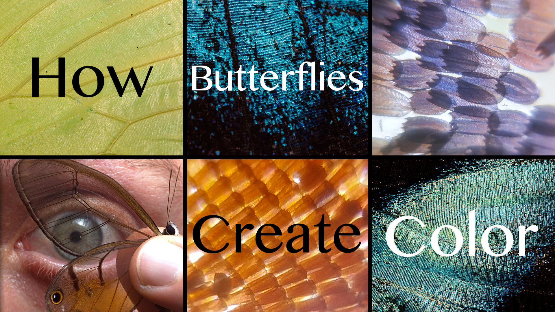
Compilation of butterfly wing scales through macro photography and Foldscope microscopy, all taken while in the field in the Amazon Rainforest.

Compilation of butterfly wing scales through macro photography and Foldscope microscopy, all taken while in the field in the Amazon Rainforest.

Hope you enjoyed, you can check out more updates via Twitter, Instagram, and YouTube!

-Aaron

Discovering a New Butterfly-Ant-Parasitic Plant Relationship

6-Butterfly-on-Tree.jpg

“Huh, that’s weird”, I muttered as I trudged through the mud in the rainforest. Even though the sun was setting, it was still hot and steamy, and sweat was dripping into my eyes as I stared at a tree with bizarre yellow outgrowths...

Watch the video "Mystery of the Yellow Bulbs: Caterpillars, Ants & Parasitic Plants"

Some sort of fungus? That was my first thought. After all, I've seen tons of strange looking fungi in the Amazon by now. But something about these yellow bulby-looking things piqued my curiosity, so I walked up to take a closer look.

A tree covered with strange yellow bulbs in the jungle

Inspecting them closer only made things more confusing. They didn't really look like fungi, at least not like any I had ever seen before. Was it a fruit produced by the tree? Plant lenticels? I started to take some pictures.

A closer look at the mysterious yellow "bulbs"

That's when I noticed something really interesting. As I scanned the tree's alien protuberances, my eyes locked on to something I wasn't quite anticipating: caterpillars! I had definitely never seen, or heard of, anything like this before. They appeared to be munching away on the yellow bulbs.

An unknown caterpillar eating one of the yellow bulbs

I quickly noticed there were also ants surrounding these caterpillars. The ants were not attacking the delicate butterfly larvae, so this had to be some sort of symbiotic relationship between the caterpillars and ants. While I had little idea at the time what I was looking at, my background in entomology was telling me one thing: this was something cool.

An ant, Ectatomma tuberculatum, guarding a caterpillar

Always gotta show a finger for scale

By this point I had returned to the tree with my colleague, wildlife photographer Jeff Cremer. We took several shots of the caterpillars, ants, and yellow bulbs. This kind of mutualistic caterpillar-ant relationship is known as myrmecophily, and has interested scientists for a long time. Caterpillars belonging to the family lycaenidae have a special structure known as the dorsal nectary organ, which secretes sugars and amino acids. This sweet, nutritious reward is what keeps the ants around and in return, the ants protect the caterpillars by driving off hungry predators and parasitoids.

An attendant ant tapping the caterpillar and receiving a nectar droplet. Filmed thanks to the help of Chris Johns

As I was inspecting this bizarre caterpillar-ant interaction, I noticed something flutter just above my head. A butterfly! Not just any butterfly, I could immediately identify it as a lycaenid and it had a distinct yellow spot on its hind wing that looked remarkably like the yellow bulbs. Was this the adult of the caterpillars!?

A butterfly with a yellow wing spot lands on the tree

Ok, Homework Time

I wrapped up my field work and headed back to the states, but I was dying to figure out what was going on here. To recap, we observed:

  • Mysterious yellow bulbs growing on a tree
  • Caterpillars eating the yellow bulbs
  • Ants taking care of the caterpillars
  • Butterfly with yellow wing spot lands on tree with yellow bulbs

I assembled the photos from the trip and starting emailing the top butterfly experts as well as botanists. The responses I received were mostly along the lines of "I've never seen anything like this before" and "there's nothing published on the life history of the butterfly". Ok, so it seemed like we were on to something new here.

With the help of some experts, we were able to identify the butterfly as Terenthina terentia, which belongs to family Lycaenidae. While this family contains around 6,000 species, the Neotropical lycaenids are still only partially known and poorly studied (Pierce 2002). Many species of lycaenids are known to engage in relationships with ants (aka myrmecophily) so our caterpillars definitely fit this criteria.

Filming this strange caterpillar-ant-parasitic plant relationship

Filming this strange caterpillar-ant-parasitic plant relationship

Several botanists emailed my pictures around to their colleagues until we were finally able to ID the yellow bulbs as a "very unusual and rarely seen" parasitic plant belonging to the family Apodanthaceae. Ever heard of that? Yeah, me either.

Apodanthaceae is a small family of parasitic plants that live inside other plants and they only become visible once the flowers burst through the bark (Bellot 2014). The species we found is possibly Apodanthes caseariae and there is little known about their ecology, what pollinates them, or how they infect their host plants. Our observations appear to be the first record of an insect utilizing Apodanthes as a host plant. The strange yellow bulbs of this plant appear to emerge once a year around October through January and then fall off.

The yellow bulbs later identified as a rare flowering parasitic plant

What's the Take-Home?

Although this species of butterfly, Terenthina terentia, was described over one hundred years ago, we knew essentially nothing about how it lived its life until now. In other words, we helped to described its life history by documenting the larval stages, host plant, and ant-associated behavior. By observing and studying this complex relationship, we can gain more insight into the diverse array of biological interactions in the Amazon rainforest.

Compilation shots of a potential Terenthina terentia caterpillar showing its morphology

However, our work is far from over and many questions still remain. Is this the butterfly's only host plant? How does it know when the parasitic plant is emerging and how does it find the yellow bulbs? What purpose does the butterfly's yellow wing spot serve? We'll attempt to continue to pick apart this fascinating Amazon mystery which will undoubtedly result in even more questions!

-Aaron Pomerantz, Entomologist

[This post originally appeared on the PeruNature.com blog http://blog.perunature.com/2015/11/mystery-of-yellow-bulbs-discovery-of.html]

You can follow Aaron for updates & get in touch on Twitter @AaronPomerantz

Special thanks to colleagues in and out of the field who assisted with this project, especially Jeff Cremer, Frank Pichardo, Chris A. Johns, Phil Torres, Christie Wilcox, Jason Goldman, Nadia Drake, Trevor Caskey, and Alex Gardels. Thanks to Andrew Warren, Alex Wild, Naomi Pierce, Adrian Hoskins, Sidonie Bellot and Robert Robbins for help with insect/plant identifications and expert input.

References

Bellot S, Renner SS (2014) The systematics of the worldwide endoparasite family Apodanthaceae (Cucerbitales), with a key, a map, and color photos of most species. PhytoKeys 36: 41-57

Pierce NE, Braby MF, Heath A, Lohman DJ, Methew J, Rand DB, Travassos MA (2002) The ecology and evolution of ant association in the Lycaenidae (Lepidoptera). Annual Review of Entomology 47: 733-771

Testing a Foldable Microscope in the Amazon Rainforest

For the past few months I've been working with Stanford University researchers who developed an origami-style paper microscope, known as the Foldscope. We were the first researchers to take the Foldscope out to the Amazon rainforest and investigate the micro-world with nothing but this incredible device and a cell phone. Below is a cross-post from www.PeruNature.com, enjoy!

A couple of months ago, I received an interesting package in the mail. It looked like a standard manila envelope, but inside was a device that could quite possibly revolutionize the way we view the microscopic world. I’m referring to the Foldscope, an origami-based optical microscope that is small enough to fit inside your pocket. The real kicker: the entire cost of the instrument is less than one dollar.

Check out our video! Foldscope in the Amazon Rainforest

The Foldscope has received some recent and well-deserved media attention (the lab’s publication on this device recently made it in the top 20 papers in PlosOne for 2014) but I hadn’t seen many videos on the Foldscope being put to the test in the field. It seemed like there was a lot of potential for this invention but I wondered how it would fare on one of my expeditions through a jungle searching for unknown species. So I decided to assemble my miniature paper microscopes and travel to one of the most remote places in the world, the rainforest of the Peruvian Amazon, to give them a go.

The Results Long story short, this device is amazing. During my time in the Amazon rainforest, I was able to investigate tiny insects, mites, fungi, and plant cells from 140x to 480x magnification without requiring a large and expensive conventional microscope.

 The cells from a flower petal recorded by connecting a cell phone to the Foldscope

Some of the diverse arthropod specimens could potentially be new to science, so it was really exciting to document images and videos of these organisms right there in the field by connecting my phone to the Foldscope.

 An unknown species of mite documented by connecting a cell phone to the Foldscope

 A spider infected by a parasitic fungus known as Cordyceps. The circles show regions of the fungus viewed under the Foldscope

In the video, I investigate bizarre structures on a plant, which are known as leaf galls. These are sometimes created by insects, but they usually have to be taken back to a lab and inspected in detail under a microscope. Lucky for me, I had the Foldscope in the field! It turns out these were in fact due to insect larvae, which burrowed into the leaf and tweaked the chemistry of the plant to produce galls. Even with macro photography, I couldn't get much detail of the larva, but at 140x magnification under the Foldscope I was able to document the morphology of this unknown critter.

 Top left, a leaf is covered in galls. Top right, a cross-section of a gall; notice the tiny insect larva living inside! Bottom, the larva was placed under the Foldscope and viewed on my cell phone. Pictures and videos were recorded in real-tim…

 Top left, a leaf is covered in galls. Top right, a cross-section of a gall; notice the tiny insect larva living inside! Bottom, the larva was placed under the Foldscope and viewed on my cell phone. Pictures and videos were recorded in real-time out in the Amazon rainforest.

Suspecting that the galls were formed by some sort of wasp or fly, I later got in touch with a couple of Diptera (fly) experts, Morgan Jackson (@BioInFocus) and Dr. Stephen Gaimari, who helped identify the gall forming culprits as a possible species of fly belonging to the family Cecidomyiidae.

Final Thoughts

The research team, led by Dr. Manu Prakash, seeks to “democratize science” by developing tools that are able to scale up to match problems in global health and science education - and I believe they are doing just that with the Foldscope. This device is cheap, easy to use, and broadly applicable whether you're a curious young student, a medical professional in the field, or someone who is interested in the numerous tiny things that surround us. Until now, I've never had a device that made viewing and sharing the microcosmos so accessible.

 Assembling the Foldscope is simple and takes less than ten minutes

 A closer look at the moss covering a tree in the tropical rainforest

 Morphology of a neotropical pseudoscorpion - all images were taken in the field

 The stinger of a honey bee viewed under the Foldscope

 I took the Foldscope with me to the beach in Santa Monica. Bottom left are kelp cells and bottom right is an unknown critter I dug out of the sand (some sort of worm?)

A breakdown of the unit costs for Foldscope components in volumes of 10,000 units, not including assembly costs (Cybulski, Clements, & Prakash 2014). The total cost of the Foldscope ranges from $0.58 to $0.97.

The Prakash lab will be starting with phase 2 of the project shortly, which will involve much larger production runs. So if you want your own Foldscope, be sure to check out their application process through www.foldscope.com and for inquiries see their contact page. Citation: Cybulski JS, Clements J, Prakash M (2014) Foldscope: Origami-Based Paper Microscope. PLoS ONE 9(6): e98781. doi: 10.1371/journal.pone.0098781

You can follow Aaron for more updates on Twitter @AaronPomerantz and the Stanford University Researchers @PrakashLab