

How does an animal become invisible? Enter the paradox of the Glasswing butterfly. As the name implies, these butterflies have transparent regions in their wings, engendering a common notion that they are “invisible” in the context of camouflage to avoid predators.
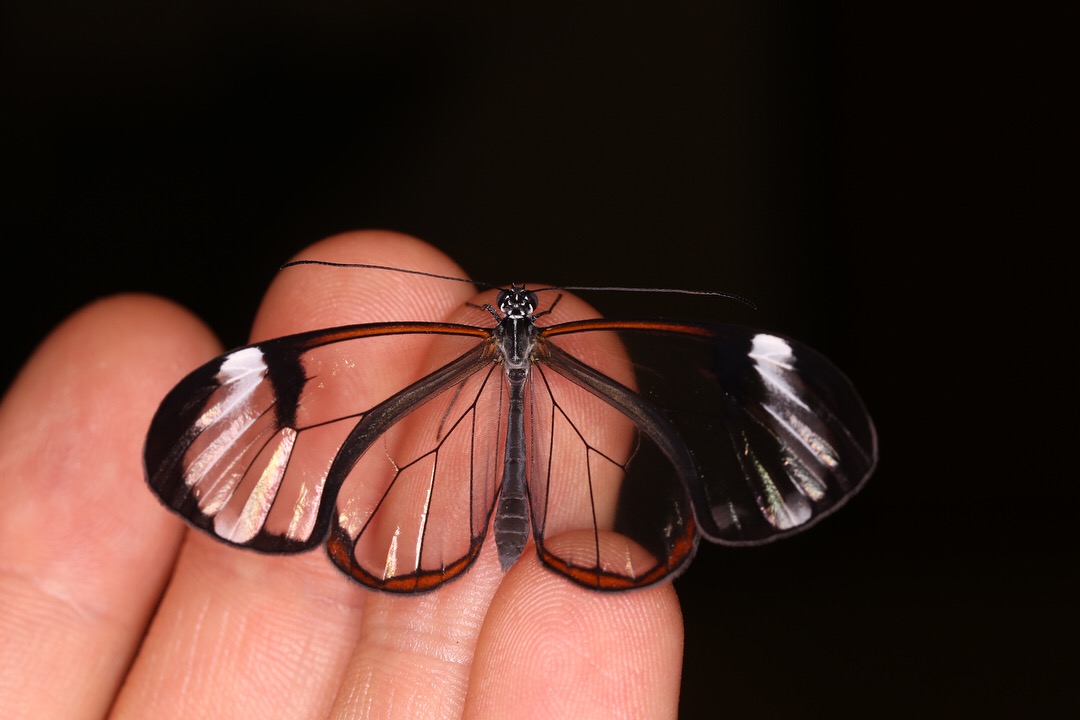
The “Glasswing” butterfly Greta oto
However, these butterflies can contain striking orange and iridescent patterns in the wings and numerous other species are known to mimic their wing patterns, highlighting the fact that these butterflies are in fact toxic, as they sequester noxious chemicals called pyrrolizidine alkaloids. Therefore, the bright colors serve as a warning signal to would-be predators such as birds.
The glasswing butterfly Greta oto was also found to harbor randomly sized nanopillars of high aspect ratio, which enables omnidirectional anti-reflection properties. More recently, another transparent-winged butterfly in the genus Chorinea was found to have dome-shaped chitin nanostructures on the wing membrane that result in wavelength-selective anti-reflective and angle-independent transmission. The aforementioned anti-reflective nanostructures found in nature are proving to be quite rewarding for applications in biomimetics and biophotonics, such as solar cells, anti-glare glasses and optical implant devices.

The role of random nanostructures for the omnidirectional anti-reflection properties of the glasswing butterfly by Siddique et al. 2015
But before diving more into transparency, let’s take a step back for a moment to consider where the color in a butterfly wing comes from in the first place. The primary unit for color in Lepidoptera is the wing scale cell and the underlying mechanism for a particular color can be due to either pigmentation from a biochemical pathway, or due to the physical architecture of scales manipulating wavelengths of light, known as structural color. To better understand processes underlying structural scale modifications, my dissertation has focused on a unique coloration strategy: wing transparency within butterflies and moths (Lepidoptera). Numerous species of Lepidoptera develop wings that allow light to pass through so that objects behind them can be distinctly seen, which has engendered a common notion that these species are “invisible” in the context of camouflage to go undetected by predators. However, my lab and collaborators hypothesize that transparency is a much more complex coloration strategy, possibly playing roles in visual communication through light polarization and iridescence. Terrestrial transparency also entails challenging optical requirements and the morphological, physiological, and genetic mechanisms involved are virtually unknown.

First visit to the Smithsonian Tropical Research Institute, Gamboa Panama
To investigate the development of transparent species endemic to the Neotropics, I realized that it was critical to obtain living specimens at various life stages. Furthermore, experiments in developmental biology often require access to tissue at precisely known time-points, and therefore being able to raise the organism of interest while preparing for trials is necessary. Therefore, I turned my sights to the Smithsonian Tropical Research Institute (STRI) located in Gamboa, Panama. The facilities at STRI look incredible, if not a touch out of place. Nestled into a small sleepy town in the rainforest, STRI has recently been upgraded to a building containing state-of-the-art molecular laboratories. This field site has also been utilized for many years by researchers investigating color pattern formation in Heliconius butterflies, who have established stellar insectaries and have knowledgeable staff on site for plant and animal husbandry.
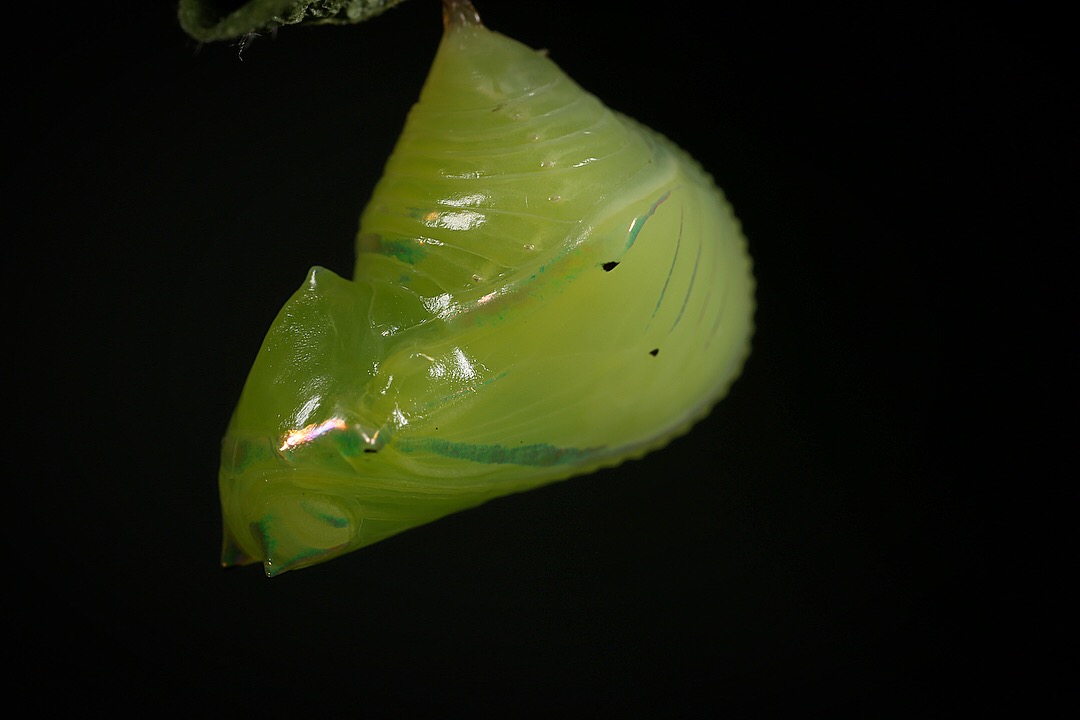
A pupa of the Glasswing raised at STRI
My goal at STRI was to raise Glassing butterflies, then investigate and experimentally manipulate pupal wings at various developmental stages in order to identify cellular and cytoskeletal scale modifications. During my expedition I was successfully able to collect and established a colony of glasswing butterflies at the local insectary. Taking advantage of the laboratory at STRI, I was able to perform dissections of pupal wings and stain wing tissue with fluorescent markers such as DAPI and phalloidin to visualize nuclei and scale cytoskeletal modifications. Additional tissue was preserved for downstream genomic and RNA experiments. Results thus far indicate that glasswing butterflies become transparent in part by modifying the ploidy levels in scale cell nuclei, as transparent regions of the wing have smaller and more spaced-out cells, and modifications of scale growth may play a role in "forked" and bristle-like scales, which allows light to pass through to the membrane of the wing which harbors anti-reflective nanostructures. This has been a critical first step to employ experiments investigating the development of transparency, including gathering material for comparative transcriptomics and functional modifications via CRISPR-cas9 genome editing. The results from this expedition and future work on the established colony can now feed into comparative analyses, help elucidate the genetic drivers of scale structural complexity, and provide insight into the evolution of terrestrial transparency.

Iridescent wing patterns at certain angles of light on the wings of transparent species
So, what’s the deal Glasswings? Are you transparent to go unseen? Are you bright to show off warning colors? Perhaps a bit of both? I think it’d be interesting if the dual nature serves to avoid a certain kind of predator under reflected light. Another possibility is that they’re showing off ultraviolet colors as warnings, which would be invisible to us, but clear as day to other animals such as birds, many of which contain opsins in their eyes capable of detecting UV. Either way, they’re a beautiful group of butterflies and it’s a beautiful scientific mystery to (attempt to) solve the evolution and development of transparency. These experiments at STRI would not have been possible without the help of the Tinker Summer Field Research Grant, to which I am very thankful for the opportunity. While this was my first visit the Smithsonian Tropical Research Institute, I have little doubt that it will not be my last.